33+ draw and label the parts of brain
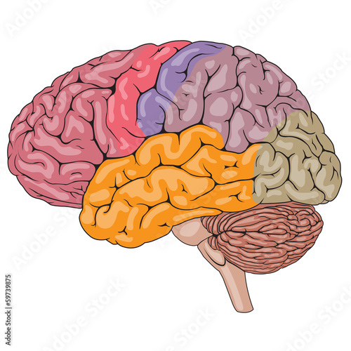
Consider a situation where a stroke or mechanical trauma has occurred resulting in damage to one of the areas of the brain indicated in the image. Right kidney 1 Urinary Bladder in Right renal artery 2 See answers Advertisement Answer 1 panesarh989 Answer.

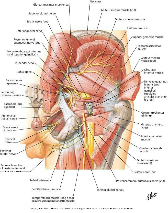
Sacrotuberous Ligament Hip Anatomy Muscles Massage Body Picture
The figure given below depicts the major and minor segments of the circle.

. This brain part controls balance movement and coordination. Drag each of the given signs and symptoms of nerve damage to the proper position to indicate the nerve most likely affected by the condition. Here you can take a quick tour of this amazing control center.
Two neurons that are passing the message along. Drag each label into the proper location in order to identify the area that would most likely. This part of the nervous system moves messages between the brain and the body.
The brain has three main parts. This brain part controls thinking. In the below-given fig.
The brain is divided into the left and right hemispheres which control opposite sides of the body. Is the largest portion of the brain encompasses about two-thirds of the brain mass - it consists of two hemispheres divided by a fissure corpus callosum it includes the cerebral cortex the medullary body and basal ganglia cerebral cortexis the layer of the brain often referred to as gray matter because it has cell bodies and synapses. The area opaca has changed into area vasculosa.
The main subcortical limbic brain regions implicated in depression are the amygdala hippocampus and the dorsomedial thalamusBoth structural and functional abnormalities in these areas have been found in depression. For each statement decide whether it is a function of the. In making such comparisons they noted that the main divisions of the human CNS the spinal cord hindbrain midbrain thalamus cerebellum and cerebrum or telencephalon were present in all vertebrates FIG.
The brains cerebral cortex is the outermost layer that gives the brain its characteristic wrinkly appearance. The brain is differentiated into fore brain mid- brain and hind brain. This brain part controls involuntary actions such as breathing heartbeats and digestion.
This is will be the cell body. Traditionally each of the hemispheres has been divided into four lobes. The brain stem is responsible for regulating the heart and lungs communications between the brain and the peripheral nervous system the nerves of the body our sleep cyc le and coordinating reflexes.
Take the two halves and twist them together into a single extension. Every second of every day the brain is collecting and sending out signals from and to the parts of your body. Cleaners of 5 different colors.
Correctly identify and label the anatomical parts of the spinal cord and its accessory structures. My art teacher taughr his entire course from this book. What Are the Parts of the Brain.
Edinger however noted that the internal organization of the telencephala showed the most pronounced differences between species. The cerebrum cerebellum and brainstem. Drag each label into the proper location in order to identify the area that would most likely have been affected.
You can see each part and later learn what areas are involved with different tasks. The human brain is the central organ of the human nervous system and with the spinal cord makes up the central nervous systemThe brain consists of the cerebrum the brainstem and the cerebellumIt controls most of the activities of the body processing integrating and coordinating the information it receives from the sense organs and making decisions as to the instructions. Primary somatosensory cortex Primary somatosensory cortex or Postcentral gyrus this is numbered rostral to caudal as 312.
You can still see some structures on the brain before you remove the dura mater. Frontal parietal temporal and occipital. The peripheral nervous system PNS is composed of spinal nerves that branch from the spinal cord and cranial nerves that branch from the brain.
Sheep Brain Dissection 1. One color each for the dendrites Any colors will do. Draw and label the following parts in the human excretory system.
The sheep brain is enclosed in a tough outer covering called the dura mater. Use arrows to show the direction of the message as it is carried along from the pre-synaptic neuron through the synapse to the post-synaptic neuron. This part of the cerebrum interprets and sorts information from the senses.
A Define excretion Name any two substances that are selectively reabsorbed. Brain The brain is composed of the cerebrum cerebellum and brainstem Fig. 2Take another pipe cleaner and attach it to the new cell body by pushing it through the ball so there are two halves sticking out.
Of Chick Embryo of 13-14 Pairs Somites or 36 Hours. Drag each label to identify the bony passageway through which the given nerve fibers pass. Each has a special function.
These two structures will likely be pulled off when you remove the dura mater. This will be the axon. Write one use of each.
A sector of a circle is the part bounded by two radii and an arc of a circle. Functional Areas of the Brain Motor Area - Control of voluntary muscles Sensory Area - Skin sensations temperature pressure pain Frontal Lobe - Movement - Problem solving - Concentrating thinking - Behavior personality mood Brocas Area - Speech control Temporal Lobe - Hearing - Language - Memory Brain Stem - Consciousness - Breathing. In doing so the brain is reduced to its parts and the whole can get lost.
Parts of a circle diagram. Keeping the body alive keeping the body balanced and allowing body systems to communicate. The kidneys are two bean-shaped organs in the renal system.
Anterior omphalomesenteric vein has developed. Important Brodmann areas Areas 123 Postcentral gyrus Gyrus postcentralis 12 Synonyms. Part of a circle bounded by a chord and an arc is known as a segment of the circle.
AOB is a sector of a circle with O as centre. Pre-Quiz Part 2. It keeps everything working even when we are sleeping at night.
The brain stem accounts for the remaining 5 of the brains mass and is along with the cerebellum the oldest part of the brain. Other major brain parts include the corpus callosum the cerebellum and the brain stem. Label the parts of the heart if youd to reference it for anatomy.
The cerebral cortex is divided lengthways into two cerebral hemispheres connected by the corpus callosum. Decreased hippocampal volumes 10 25 have been noted in subjects with depression. Our free diagrams and quizzes on the parts of the brain are a great place to get started.
Take special note of the pituitary gland and the optic chiasma. The important things to consider here are which parts of your business generate trust and which parts generate utility.

Fox Outline Drawing At Paintingvalley Com Explore Collection Of Fox Outline Drawing Animal Coloring Pages Outline Drawings Fox Coloring Page

Pin By Taina Rainele On Art Drawing Design Anatomy Art Art Tattoo Brain Tattoo

Opening The Flowers Of The Lungs Via The Pelvis Engaging In Biointelligent Somatic Yoga Practices To Celebrate The Spri Human Anatomy Art Skull Art Medical Art

1 622 Thalamus Human Health Wall Murals Canvas Prints Stickers Wallsheaven

Tattoo Idea Tattoo Ideas Central Heart Painting Half Sleeve Tattoo Heart Tattoo

Flowers Doodles Pattern Simple 33 Ideas Flower Pattern Drawing Flower Doodles Tattoo Design Drawings
Off Topic Limbic System And Brain Can Both Be Retrained Plasticity March 2019 Vitamindwiki

Pin On Risunki

Pin Em We All Like Shop
Variety Of Evidence That Vitamin D Helps The Brain July 2014 Vitamindwiki

Human Brain Vector Illustration Labeled Anatomical Educational Parts Scheme Wall Mural Vectormine

Botany Chloroplasts And Stroma Video Organic Chemistry Introduction To Organic Chemistry Chemistry

8 657 Brain Cerebellum Cerebra Wall Murals Canvas Prints Stickers Wallsheaven

I Wonder If We Could Find A Middle Ground Between Flat Illustration And Something More Realistic Like This Lungs Art Drawings Anatomy Art

The Human Brain Is Side View Illustration Wall Mural Wallpaper Murals Vasilisatsoy